
Unveiling the Contrasts
BF and DF Imaging, Reflected and Transmitted Illumination


In the world of microscopy, imaging techniques play a crucial role in revealing the intricate details of samples. Two commonly used imaging methods are Bright-field (BF) and Dark-field (DF) microscopy, which utilizes different illumination techniques to enhance contrast. Additionally, microscopy techniques can employ either transmitted or reflected illumination, further expanding the range of imaging possibilities. In this blog post, we will explore the differences between BF and DF imaging and shed light on the concepts of reflected and transmitted illumination.
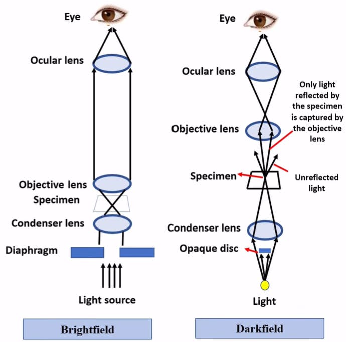
Figure 1: Schematic difference between BF and DF
BF Imaging: Illuminating the Path to Clarity
Bright-field imaging is one of the simplest and most widely used techniques in microscopy. In this method, the sample is illuminated with a uniform and direct light source, typically from below the sample, and the transmitted light passes through the specimen and reaches the objective lens. The bright-field microscope utilizes a condenser to focus the light onto the sample and an objective lens to magnify the transmitted light, forming the final image.

Figure 2: Bright-field illumination, sample contrast comes from the absorbance of light in the sample.
BF microscopy provides high contrast between the sample and the background, as the majority of light passing through the specimen is absorbed or scattered, leading to dark regions in the image. This technique is excellent for observing stained samples or those with inherent contrast, such as cells in tissue culture or stained microorganisms. However, it may lack contrast when imaging transparent or low-contrast specimens.
DF Imaging: Illuminating the Unseen
Dark-field microscopy is designed to enhance the visualization of transparent or low-contrast samples by manipulating the illumination. In DF imaging, the light is directed at an oblique angle, so it bypasses the objective lens and does not enter directly into the microscope's image-forming path. Instead, only the light scattered or refracted by the sample reaches the objective lens, producing a bright image against a dark background.

Figure 3: Dark-field illumination, sample contrast comes from light scattered by the sample.
DF microscopy is particularly useful for samples with refractive index variations, such as live cells, microorganisms, or small particles. It enhances the visualization of fine structures and small details that might be challenging to observe using other techniques. By capturing scattered light, DF imaging brings out the subtle features that would otherwise go unnoticed in bright-field microscopy. Basically, the smaller the sample, the better it is to use dark field imaging. DF imaging can be upgraded from BF imagining by simply placing an opaque filter between the light and lower objective lens below your sample!
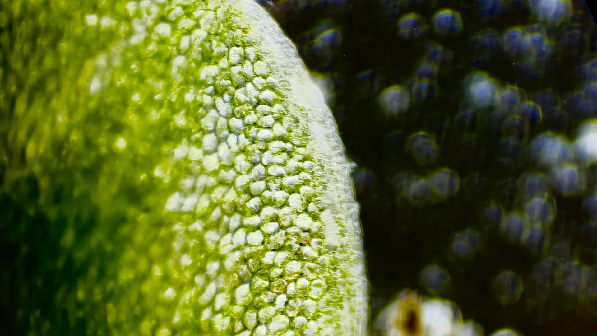
Figure 4: DF Image of plant organ
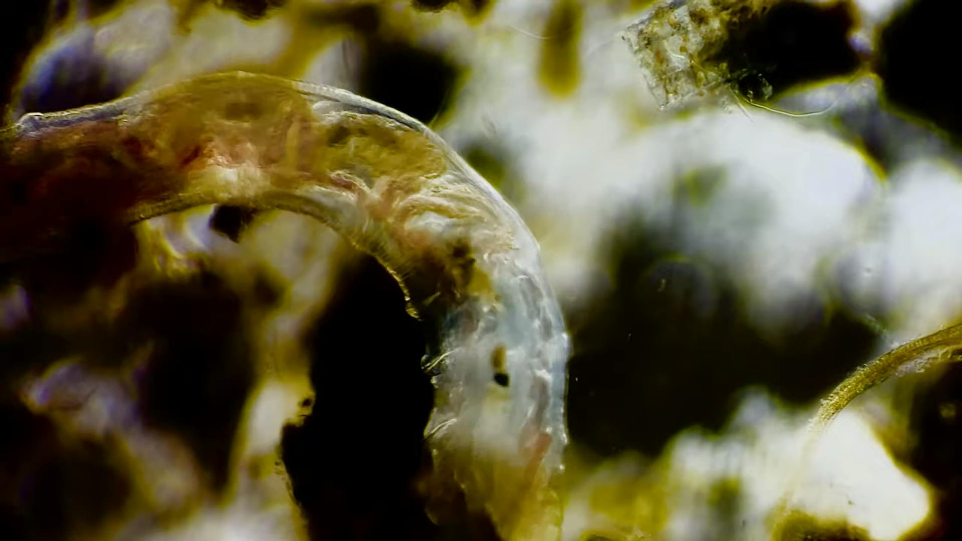
Figure 5: DF Image of worm species eating biomatter
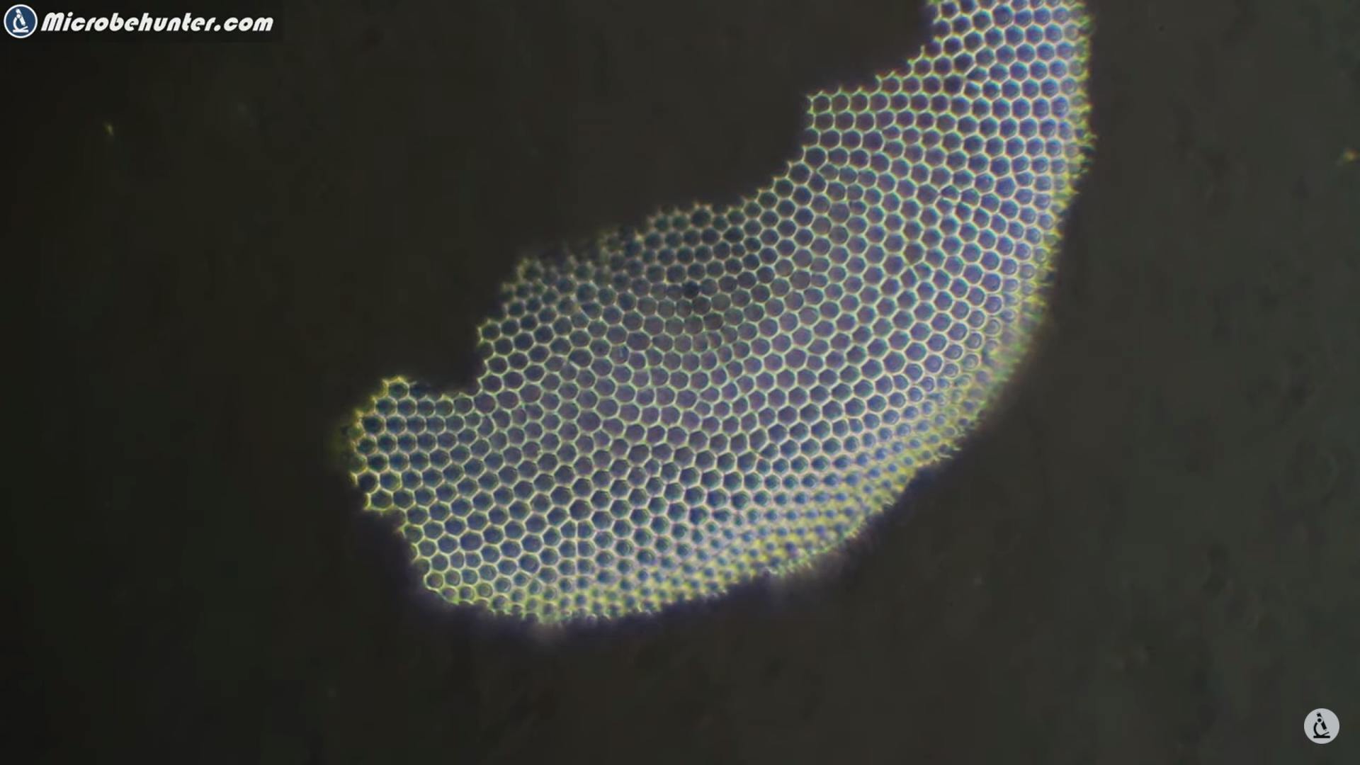
Figure 6: Marine worm diatoms
Reflected Illumination: Shining a Light on Surface Details
In microscopy, reflected illumination is employed when examining opaque samples or those with surface structures. This technique involves illuminating the sample from above, and the light is reflected off the surface and captured by the objective lens. Reflected illumination can be achieved using various light sources, such as halogen lamps or LEDs, and can be enhanced by the use of specialized filters.
Reflected illumination is particularly suitable for studying metal surfaces, geological samples, or materials with topographical features. It provides valuable insights into surface morphology, texture, and the distribution of reflective or opaque elements. By manipulating the angle and intensity of the incident light, researchers can further enhance contrast and highlight specific surface details.
Transmitted Illumination: Peering through the Inner Depths
Transmitted illumination is the standard method used in most microscopy techniques, including BF and DF imaging. This technique involves passing light through the sample, allowing the observer to visualize internal structures or features. The transmitted light can be absorbed, scattered, or refracted by the sample, providing information about its composition and organization.
Transmitted illumination is widely applicable and can be utilized for various samples, including thin sections of tissues, biological cells, or transparent materials. By employing different contrast-enhancing techniques, such as phase contrast or differential interference contrast (DIC), the visibility of cellular structures or subcellular components can be significantly improved.
Conclusion
BF and DF imaging techniques, along with reflected and transmitted illumination, offer distinct advantages for microscopy applications. Bright-field imaging provides excellent contrast for stained or high-contrast samples, while dark-field imaging enhances the visibility of transparent or low-contrast specimens. Reflected illumination allows for surface analysis, highlighting surface morphology and reflective features, while transmitted illumination reveals the internal structures of samples.
By understanding the differences between BF and DF imaging, as well as the concepts of reflected and transmitted illumination, scientists can select the most appropriate technique for their specific research needs. With these imaging tools at their disposal, researchers can delve deeper into the microscopic world and uncover new insights across various scientific disciplines.