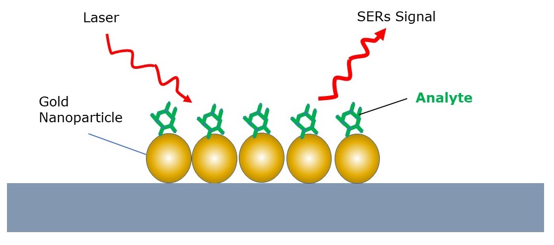
In Raman spectroscopy, SERS has been a common method to enhance the Raman scattering signal of materials which are low in the Raman active analytes. However, this requires the the analyte to be absorbed onto rough metal surfaces or nanoparticles.
What is SERS?
More specifically, this enhancement is either through electromagnetic or chemical mechanisms.
When light interacts with the rough metal surface or nanostructures, it can excite coherent surface electron oscillations. These electron oscillations create strong electromagnetic fields at the metal surface. Molecules near the surface experience this enhanced electromagnetic field, leading to a significantly increased Raman signal through this feedback mechanism.

Moreover, the chemical enhancement is due to charge transfer between the adsorbed molecule and the metal surface. The interaction can result in an increase in the polarizability of the molecule, thereby enhancing the Raman scattering.
SERS is great!...when you don't mind 'gluing' nanoparticles to your sample. What if you have a sensitive sample? What if you don't want any surface or particle to contaminate it?
Can't we just make the instrument better?
Instead of applying surfaces and nanoparticles to our sample, we can enhance our signal by making a few instrumentation design updates. In this article, we'll evaluate a few probe designs which are geared towards this enhancement of liquid samples.
The Confocal Raman Microscope
The standard confocal Raman microscope consists of an excitation laser: providing the monochromatic light required for Raman scattering; laser line filter: ensuring that only the desired wavelength is directed towards the sample, removing any unwanted laser drifts. A beam splitter not only directs the laser light towards the sample but also allows the scattered light to pass through to the spectrometer. Objective lenses focus the laser light onto a small spot on the sample and collects the back-scattered light. High numerical aperture lenses are often used to achieve better spatial resolution and signal collection since they collect a wider angle of the isotropic Raman scattering sphere. The biggest advantage of any microscope is the precision its motorized XYZ stage: this allows precise positioning in three dimensions (x, y, and z), facilitating detailed chemical mapping of the sample.

When the back-scattered light passes back through the dichroic edge filter, it selectively blocks the strong Rayleigh scattered light and transmit the weaker Raman scattered light, ensuring that the spectrometer detects only the Raman signal. Furthermore, a notch filter is placed to block more Rayleigh scattered light. A confocal pinhole is placed at a conjugate focal plane to the sample, it blocks out-of-focus light, improving spatial resolution and reducing background noise. This Raman scattered light passes to the spectrometer which consists of a diffraction grating: this physically disperses the collected scattered light into its constituent wavelengths. The physically dispersed light can be mapped onto a detector, typically a Charge-Coupled Device (CCD) that captures the dispersed light and converts it into an electrical signal for analysis. The CCD detector is often cooled to reduce thermal noise.
Axicon Lens
Liquids are often stored in glass vials, meaning the sole viable option for conducting Raman analysis of a bioreactor is to pass a laser through a window separation. Therefore, this presents the problem of exciting fluorescence transitions within the glass which interferes with the Raman spectrum. Nonetheless, the use of an axicon lens has shown promise in Spatially Offset Raman Spectroscopy (SORS) and has been a central component for reducing the laser flux through glass windows, allowing the analyte within the container to be analysed with a reduced fluorescence spectrum. H Fleming et al. [a] demonstrate this application for the analysis of whisky through the bottle.


Elliptical Silver Satellite
Some lasers are very difficult to completely collimate (in fact its actually impossible). In my undergraduate work, developing a portable Raman device, the collimator we used didn't actually completely collimate the beam, so I had to assume it was a concave lens when I was applying thin lens equations to find the focal point of my Raman excitation spot. A similar problem occurs in all Raman devices, particularly those whereby the beam travels a distance larger than 10cm. How do we combat this divergence?

Communication signals also diverge. Electromagnetic waves used for sending communication signals are received using large elliptical satellite dishes which focus the signal onto the central receiver, the waves can enter the dish at any orientation and still become focussed onto the receiver.
The same may be applied to a laser which diverges in orientation when attempting to collimate. Let the laser pass through the liquid media you wish to analyse (hoping that no signal will be lost on the way), from there the multiple diverging beams can focus on the focal point of the elliptical mirror and induce Raman scattering at this focal point. Raman scattering occurs isotopically, therefore, the bottom scattering sphere will assemble onto the receiving dish, reflect off the mirror and pass through the media along the same path as the initial laser. Likewise, the top scattering sphere will pass through the media, however, will be diverged into a sphere.

There may exhibit some interreference from the top and bottom spheres since there is a path difference between the Raman light. This setup requires a further set of large optics to capture, filter and focus the light into the spectrometer, assuming that path differences, low flux densities as well as spurious scattering does not disrupt the Raman signal.
Gold Backscattering Mirror
Demonstrated by M. Lui [b], this is similar to the Elliptical setup, but isolates laser light which passes through the focal point and is wasted to the media. Instead of wasting this laser light, an hyperbolic gold mirror may be placed at the bottom of the container and enhance the Raman scattering by adding an additional excitation to the laser spot.

This method has been shown to enhance the Raman scattering intensity by up to 6x as much as without the mirror. I hope to use this method in my own work with regards to bioprocesses, whereby an immersion probe can have a gold mirror attachment immersed into the bioreactor, which can reflect the diffused laser light, and enhance the signal through backscattering enhancement. The biggest issue is that bioproducts are not to be touched unless they are FDA approved sensors. Reaching FDA approval would take up to 5 years, by which point another engineering firm has made a new design and has overtaken the approval process.
Freeform Optics
One of the most interesting assemblies is the use of freeform optics, whereby the reflection of optical surfaces is minimised by passing the light through a single optical component which totally internally reflects the light such that no Raman or laser light is lost in the process. These freeform optics have been 3D printed using the additive manufacturing methods, VeroClear, a glass material, printed by the Objet30 Pro, and has shown to collect adequate Raman scattering.


However, this investigation concluded that there is no enhancement, but rather a significant reduction in Raman intensity, demonstrating that this is method poses no advantage to the optical components. I believe a better material could have been used, with other materials instead of reflective surfaces and optically inactive surfaces. A method similar to the creation of standard optics, with the heating of glass would make the freeform optic more suitable as it would be similar to standard optical components. scattering light may have been lost due to spurious scattering from within the material.
Can you make it better?
Sure you can. With some investigative abilities and scientific demonstrations, we can engineer something to enhance our Raman scattering signal - without the use of SERS.
References
[a] FLEMING, H., CHEN, M., BRUCE, G. D. & DHOLAKIA, K. 2020. Through-bottle whisky sensing and classification using Raman spectroscopy in an axicon-based backscattering configuration. Analytical Methods, 12, 4572-4578.
[b] LIU, M., MU, Y., HU, J., LI, J. & ZHANG, X. 2021. Optical Feedback for Sensitivity Enhancement in Direct Raman Detection of Liquids. Journal of Spectroscopy, 2021, 5588417.
